


About us
Cardisio Türkiye is the brand that offers Cardisiography to physicians and clinics in Türkiye. It is operated by CardiCare, distributor for Türkiye and the Turkic Republics.
Who we are

Cardisio Türkiye speaks about AI-supported heart screening in a clinical voice that rests on evidence and does not exaggerate. CardiCare carries the warm, human face of the same service.
Distributor

CardiCare is the distributor of Cardisio GmbH for Türkiye and the Turkic Republics. Our legal entity is Cardicare Medikal Sağlık ve Danışmanlık Hizmetleri Sanayi ve Ticaret Limited Şirketi.
Manufacturer: Cardisio GmbH
Cardisio is a private mobile health company headquartered in Frankfurt, Germany. It works on decision support for the early detection of ischemia in coronary artery disease. By screening more people, faster and with higher accuracy, it aims to sharply reduce medical costs, loss of quality of life and deaths. To this end it has developed a screening test called “Cardisiography”.
Cardisio GmbH was founded in 2016. Cardisiography is now used in a multitude of practices and clinics worldwide.
Vision
In Cardisio's future, everyone has access to a scientifically proven, highly accurate and non-invasive heart screening procedure that is easy to use, examiner-independent and affordable.
Method
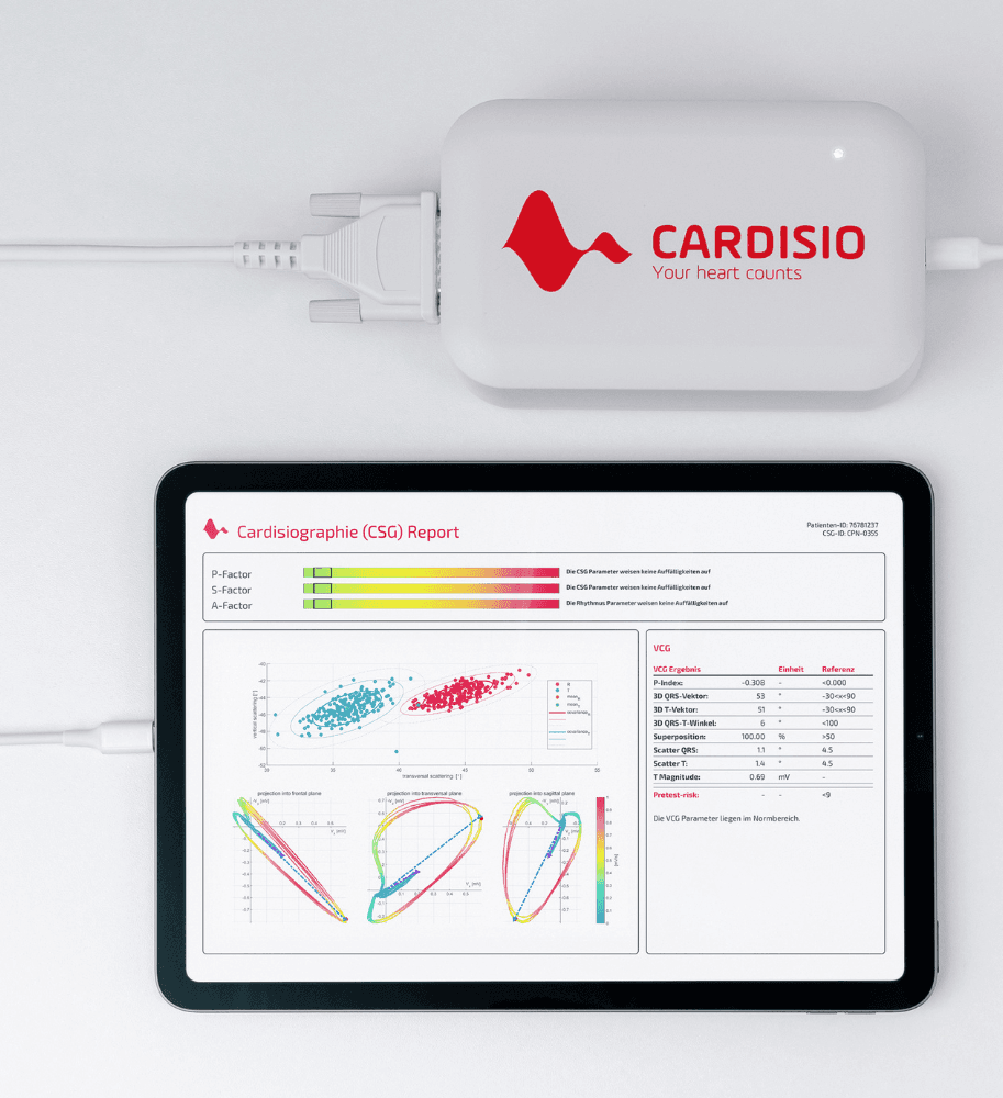
As a method, Cardisiography is based on vectorcardiography (VCG), which performs a 3-dimensional scan of the heart's electrical activity. The measurement is taken with 4 electrodes placed on the front of the chest and 1 electrode placed on the back. After the vectorcardiogram has been recorded with the 5 electrodes, a 12-lead ECG is also obtained by mathematical transformation. Using artificial intelligence and machine learning, the algorithm analyzes both VCG and conventional ECG parameters. The Cardisio algorithm processes 3,200,000 data points per measurement. After analyzing 290 different VCG and ECG parameters, it presents the user with a final report.
The report provides a set of ECG and VCG parameters, each of which the literature has shown to be of great value in predicting cardiac events. It also provides 3 separate bars, called the P factor, S factor and A factor, shown in different colours according to a risk scale. The P factor is positioned as a predictor of perfusion disorder caused by ischemia, the S factor of the presence of structural heart disease, and the A factor of the presence of arrhythmia.
The recording takes about 4 minutes, and the report is ready within 1 minute of the recording. It is a non-invasive, fast screening test. As the analysis is operator-independent, it eliminates operator-related error in the analysis. No stress induction is needed during the measurement. This makes Cardisiography more practical than the stress-based tests we use to detect coronary ischemia, such as the exercise test and MPS, and it can also be performed on patients who cannot take an exercise test.
Literature and validation
The literature has shown VCG to be superior to the ECG in many areas, such as predicting atrial and ventricular enlargement (1), detecting electrically inactive areas (2), correlation with echocardiography (3), detecting myocardial infarction in bundle branch block (4), diagnosing Brugada syndrome (5), and locating the accessory pathway and ventricular pre-excitation in pre-excitation syndromes (6). Because the Cardisiography algorithm is built not only on the conventional ECG but on the combination of ECG and VCG, its decision support is expected to show high sensitivity, specificity and accuracy.
Likewise, in the validation study of Cardisiography for the detection of coronary artery disease, sensitivity was 90.2 ± 4.2% in women and 97.2 ± 3.1% in men, specificity was 74.4 ± 9.8% in women and 76.1 ± 8.5% in men, and overall accuracy was 82.5 ± 6.4% in women and 90.7 ± 3.3% in men (7).
Our brand essence
Technology that listens to the heart, care that sees the person.
Where to find us
You will find our address and contact details below. You are always welcome to write to us with your questions.
Türkiye and the Turkic Republics
Cardicare Medikal Sağlık ve Danışmanlık Hizmetleri Sanayi ve Ticaret Limited Şirketi
Küçükbakkalköy Mh. Dudullu Cad. Brandium Rezidans R1 Blok No:23-25A, K:12 D:141-142, Ataşehir/İstanbul
Centres offering the test
Find out moreCentres offering the test
References
- Chou TC. When is the vectorcardiogram superior to the scalar electrocardiogram? J Am Coll Cardiol 1986;8:791–799
- Pipberger HV, Goldman MJ, Littmann D, Murphy GP, Cosma J, et al.: Correlations of the orthogonal electrocardiogram and vectorcardiogram with constitutional variables in 518 normal men. Circulation 1967;35:536–551
- Bocanegra Arroyo J, Braga JMS, Luna Filho B. Análise crítica do eletrocardiograma e do vetocardiograma no diagnóstico da hipertrofia ventricular esquerda. Rev Soc Cardiol Estado de São Paulo 1994;4:353–360
- Hurd HP II, Starling MR, Crawford MH. Comparative accuracy of electrocardiographic and vectorcardiographic criteria for inferior myocardial infarction. Circulation 1981;63:1025–1029
- Atarashi H, Ogawa S, Harumi K. Idiopathic Ventricular Fibrillation Investigators. Three-year follow-up of patients with right bundle branch block and ST segment elevation in the right precordial leads: Japanese Registry of Brugada Syndrome. Idiopathic Ventricular Fibrillation Investigators. J Am Coll Cardiol 2001;37:1916–1920
- Giorgi C, Nadeau R, Primeau R. Comparative accuracy of the vectorcardiogram and electrocardiogram in the localization of the accessory pathway in patients with Wolff-Parkinson-White syndrome: validation of a new vectorcardiographic algorithm by intraoperative epicardial mapping and electrophysiologic studies. Am Heart J 1990;119:592–598
- Braun T, Spiliopoulos S, Veltman C, Hergesell V, Passow A, Tenderich G, et al. Detection of myocardial ischemia due to clinically asymptomatic coronary artery stenosis at rest using supervised artificial intelligence-enabled vectorcardiography - A five-fold cross validation of accuracy. J Electrocardiol. 2020;59:100-5. On the clinical evidence page